

Back and leg pain that gets worse when you stand or walk, then eases when you sit down, is a frustrating pattern. For many adults, especially those over 50, it points to a condition called spinal stenosis. It can make simple activities like grocery shopping or a walk around the block feel exhausting.
Getting the right diagnosis is the first step toward relief. Yet stenosis can be tricky to capture on imaging, because the symptoms often depend on body position. This is where the way an MRI is performed can make a real difference.
What Is Spinal Stenosis?
Spinal stenosis is a narrowing of the spaces inside the spine. This narrowing can put pressure on the spinal cord or the nerves that branch out from it. It most often affects the lower back, known as lumbar stenosis, and the neck, known as cervical stenosis.
The narrowing usually develops slowly over time. Common causes include age-related wear and tear, thickened ligaments, bulging or herniated discs, and bone spurs from arthritis. In some people, a vertebra slipping slightly forward over the one below it, called spondylolisthesis, also reduces the available space.
Common Symptoms to Watch For
Lumbar stenosis often causes pain, numbness, tingling, or weakness in the lower back, buttocks, and legs. A classic sign is leg discomfort that builds while walking or standing and improves when sitting or leaning forward. Many people notice they feel better leaning on a shopping cart.
Cervical stenosis tends to cause neck pain along with numbness or weakness in the arms and hands. In more advanced cases, it can affect balance and fine motor skills, such as buttoning a shirt. Any sudden loss of bladder or bowel control is a medical emergency and needs immediate care.
Why Position Matters in Stenosis
The spine is not a fixed structure. It changes shape as you move. When you stand, gravity compresses the spine and the discs bear your full body weight. When you bend backward, the spinal canal naturally narrows. When you bend forward or sit, it tends to open up.
This explains why stenosis symptoms come and go with posture. It also explains a key challenge in diagnosis. If imaging is done only while you lie flat and relaxed, the spine is in its least stressed position, and the narrowing that causes your symptoms may look milder than it really is.
The Limits of a Traditional Lying-Down MRI
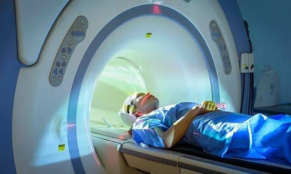
A conventional MRI is performed with the patient lying on their back inside a closed tube. It produces excellent detail and remains a valuable tool. However, lying down takes weight off the spine, which can reduce disc bulging and ease pressure on the nerves during the scan.
As a result, some patients receive results that do not seem to match how severe their pain feels in daily life. A scan may show only mild narrowing, while the patient struggles to walk more than a short distance. This mismatch can lead to confusion, delayed treatment, or repeated testing.
How Upright MRI Changes the Picture
Upright MRI allows the spine to be scanned in a weight-bearing position, such as standing or sitting. Some systems also allow imaging while the patient bends forward, bends backward, or rotates. This lets doctors see the spine under the same conditions that trigger symptoms.
Because the spine is carrying body weight during a weight-bearing upright MRI, narrowing that only appears under load may become easier to see. Disc bulges, ligament thickening, and slippage between vertebrae can look different when the patient is upright compared with lying down.
Imaging in the position of pain gives physicians a fuller understanding of what is happening. It can help connect what the patient feels with what the images show, which is the foundation of a confident diagnosis.
A More Comfortable Experience for Many Patients
Many people with stenosis find it painful to lie flat for an extended period. Others feel anxious or claustrophobic in a closed MRI tube. Both issues can make it hard to stay still, and movement during a scan can blur the images.
Open upright MRI systems are designed with open sides, so patients can see around them and often feel more at ease. Being able to sit or stand in a more natural position can make the scan more tolerable, which helps produce clearer results.
How Better Imaging Shapes Treatment
An accurate picture of stenosis helps doctors choose the right path forward. Mild cases may respond well to physical therapy, posture changes, activity adjustments, and medication. Some patients benefit from epidural steroid injections that reduce inflammation around the nerves.
When conservative care is not enough, surgery may be considered. Procedures such as decompression create more room for the nerves, and in some cases, spinal fusion is used to stabilize the spine. Clear imaging helps surgeons understand exactly where the compression is and how it changes with movement, which supports careful planning.
Questions to Ask Your Doctor
If you suspect stenosis, talk with your doctor about how your symptoms change with standing, walking, and sitting. Ask whether positional or weight-bearing imaging could add useful information in your case.
If you have already had a lying-down MRI that does not seem to explain your symptoms, it is worth asking whether an upright scan might help. Your doctor can decide which type of imaging best fits your history and goals.
Conclusion
Spinal stenosis is closely tied to posture and movement, which can make it hard to capture on a traditional lying-down scan. By imaging the spine while it bears weight, upright MRI can reveal narrowing that might otherwise be underestimated and help connect symptoms with clear findings.
For patients whose pain does not match their past results, or who struggle with closed scanners, learning about the benefits of upright MRI can be a helpful first step toward a clearer diagnosis and a treatment plan that fits.
